
The Common vein Copyright 2011
Introduction
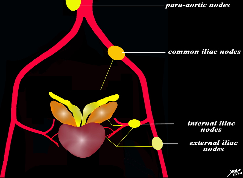
Lymphatic drainage from the prostate is via the internal iliac, sacral, vesical, and external iliac nodes.
|
Lymphatic Drainage |
|
The lymphatic drainage from the prostate is accommodated by the external iliac nodes, internal iliac nodes which will both drain to the common and paraaortic systems. Courtesy Ashley Davidoff Copyright 2011 All rights reserved 96269b35b01d01e02L.8sp01 |
Applied Biology – Iliac Nodes
|
Nodal Metastases |
|
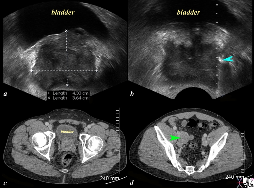
The series of US and CT scans are from a 50 year old man with an elevated PSA Image (a) is a transverse ultrasound showing a minimally prominent nodular prostate. Image b shows the needle tip (teal blue arrow) in the left peripheral zone. Image c is a transverse image of the prostate showing a nodular anterior surface while image d shows an almost 3cms external iliac node. The biopsy was positive for prostate carcinoma Image Courtesy Ashley Davidoff MD Copyright 2010 97374c.81s |
|
Local Spread and Iliac Nodes |
|
The CT scan of the pelvis is from a 88 year old man with known prostate carcinoma. Image a and b are through the bladder (yellow) showing the abnormal prostatic gland (green) which is growing toward the left side. A stent (solid white tube traverses the prostate and a second tube with hollow center represents the tip of a Foley catheter in the bladder. The seminal vesicles (pink) are seen posteriorly as symmetric structures except for mild compression of the left by the tumor. Image c and d reveal an almost 2cms left internal iliac node through which the stent passes as well. Its size and participation in the obstruction of the left ureter is incriminating. Image Courtesy Ashley Davidoff MD Copyright 2010 25481c01.8s |

Retroperitoneal
Local Extension and Retroperitoneal Adenopathy |
|
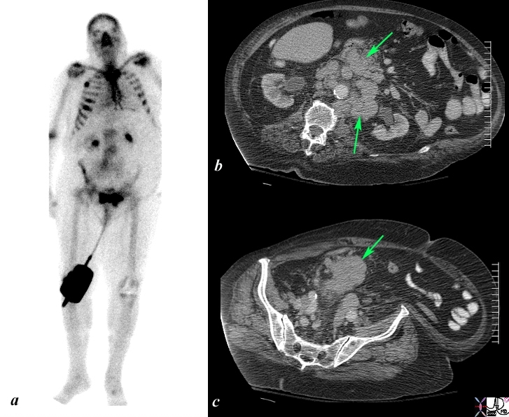
The Bone scan and CT scan is from a 77year old man with known prostate carcinoma. Image a shows multiple hot spots in the ribs consistent with blastic metastatic bone disease. Image b shows extensive lymphadenopathy (green arrows) in the retroperitoneum of the upper abdomen surrounding the ureters resulting in early mild hydronephrosis. . Image c shows a recurrent peritoneal mass (green arrow). These findings are consistent with metastatic prostate carcinoma in the peritoneal cavity, retroperitoneal nodes, and ribs. Image Courtesy Ashley Davidoff MD Copyright 2010 98965c.8s |