Copyright 2010
Introduction
Normal Reference
|
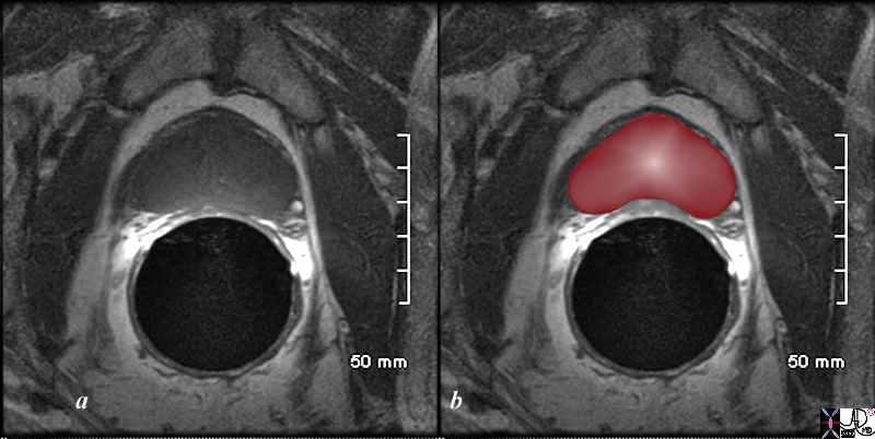
Heart Shaped in the Transverse Plane T1 Weighted MRI |
|
The patient is a 60 year old man. His MRI was performed with a transrectal coil and the image shows the T1 weighted sequence in the axial projection (a,b). The scan shown demonstrates the normal heart shape of the prostate at this level in the axial projection. Zonal anatomy cannot be distinguished since the T1 weighted sequence results in a homogenous signal from all the zones. Image Courtesy Ashley Davidoff MD Copyright 2010 98874cL.8s |
BPH Smooth Lobular
|
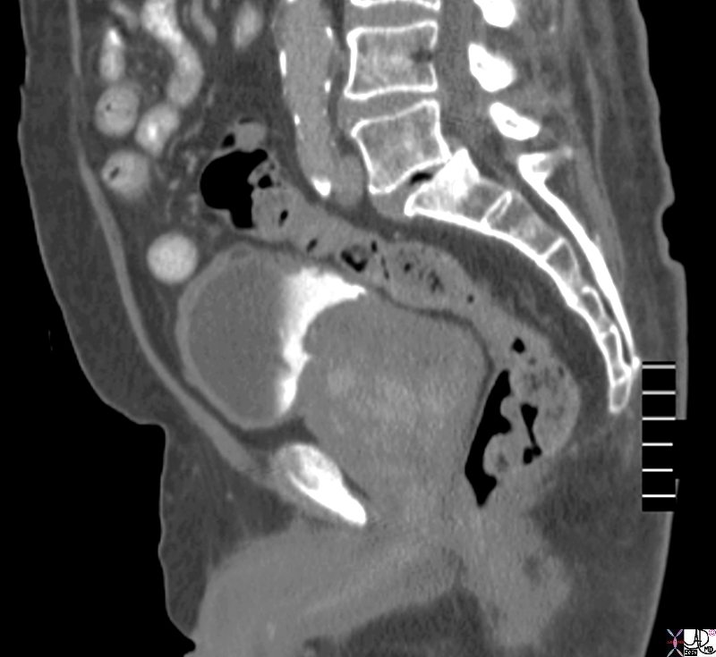
Large Smooth Gland in the Sagittal Plane |
|
The CTscan reconstructed in the sagittal plane is from a patient with an enlarged prostate. The anterior aspect of the gland protrudes with two symmetrical homogeneous components likely representing an enlarged transitional zone and prostate hyperplasia though by the nature of the disease the combination of cancer and BPH commonly coexist. The matrix of the gland is heterogeneous The bladder wall is thickened anteriorly. Image Courtesy Ashley Davidoff MD Copyright 2010 76771.8s |
|
Large Bilobed Smooth Gland |
|
The CTscan is from a patient with an enlarged prostate. The anterior aspect of the gland protrudes with two symmetrical homogeneous components likely representing an enlarged transitional zone and prostate hyperplasia though by the nature of the disease the combination of cancer and BPH commonly coexist. The urinary bladder wall is minimally thickened. Image Courtesy Ashley Davidoff MD Copyright 2010 25517b.8s |

Irregular Gland
|
Large Irregular Gland |
|
The CTscan is from a patient with an enlarged prostate. The anterior aspect of the gland protrudes into the bladder with an irregular shape (green arrow). The bladder wall is slightly thickened (orange arrow). The peripheral zone is suggested by a hypodense posterior region (maroon arrow) Image Courtesy Ashley Davidoff MD Copyright 2010 24842.81s |

Cancer Irregular
|
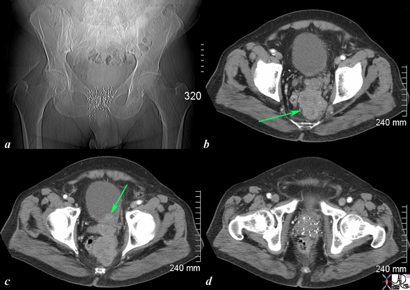
Irregular Shape of Carcinoma as it Invades into Surrounding Tissue |
|
The CT scan of the pelvis is from an 76 year old man with known prostate carcinoma treated with implanted radiation seeds. Image a shows multiple radiation seeds within the prostate gland Image b (light green arrow) shows an irregular heterogeneous mass measuring about 5cms extending above the treated field and progressing posteriorly to the presacral space. . Image c shows the same mass as b extending into the base of the bladder (green arrow). Image d shows multiple prostatic seeds in the prostate gland. These finding are consistent with prostate carcinoma treated with implanted radiation seeds but with the tumor now continuing to grow superiorly beyond the confines of the treatment, and inward to the base of the bladder. . Image Courtesy Ashley Davidoff MD Copyright 2010 98929c01.8s |