The central zone has both glandular elements and ductal elements and characteristically surrounds the ejaculatory ducts and stromal elements. The epithelial cells are slightly different and are composed of tall columnar cells with eosinophilic cytoplasm, and prominent basal cell layer.
|
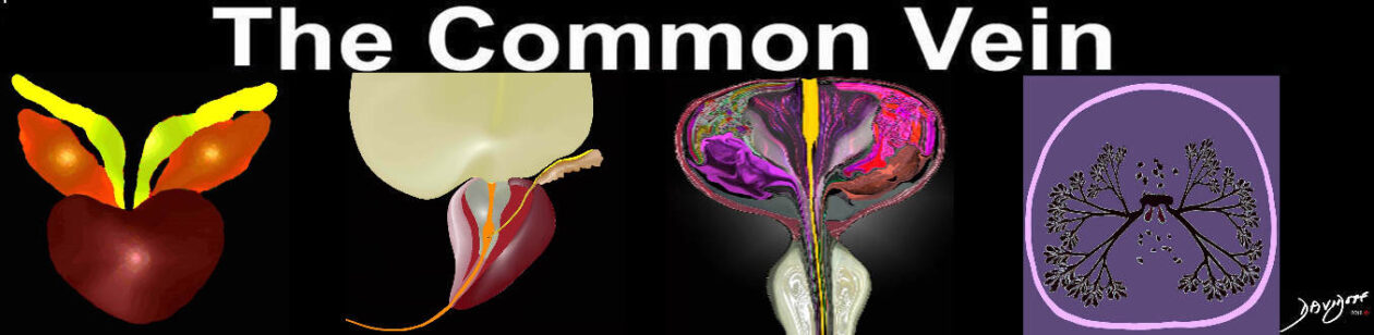
The Central Zone – A Different Histological Makeup |
|
The diagram reflects the basic histological makeup of the prostate in axial projection with a focus on the central zone (intermediate pink. The glandular elements are a minor component of this zone and they are different in nature compared to the epithelial elements of the peripheral zone, while the stromal elements are more prominent in this region functioning as the required force for expulsion of secretions during ejaculation. The central zone incorporates the ejaculatory ducts (yellow). Courtesy Ashley Davidoff Copyright 2011 42707b03b45b050prb04a.8s |
|
Zonal Anatomy of the Prostate in Transverse Plane |
|
The prostate gland can be viewed as having 4 major zones. In the young adult male the outer zone called the peripheral zone, accounts for 70% of the parenchyma. Inward of the peripheral zone is the central zone that accounts for 20% of the parenchyma. The periurethral zone is called the transitional zone and fibromuscular layer (anterior layer) share the remaining 10% of parenchyma Courtesy Ashley Davidoff MD Copyright 2010 25078b04b02L05L.8s |

|
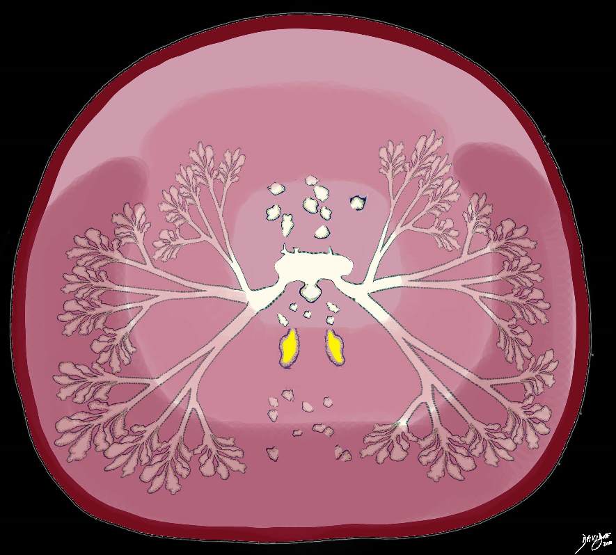
The Prostate in Sagittal View |
|
The diagram reveals the prostate in sagittal view. The peripheral zone is the largest portion and occupies the base, extending the entire length of the posterior wall to the apex. The central zone lies inward of the peripheral zone and is that part through which the ejaculatory duct courses. The transitional zone surrounds the urethra, and the anterior fibromuscular layer runs from the base to the apex anteriorly. The ureter is overlaid in orange and the ejaculatory duct empties into the widened portion of the urethra called the verumontanum Courtesy Ashley Davidoff MD Copyright 2010 99653b11b04L.8S |
|
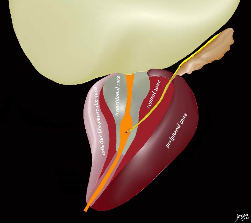
Zonal Anatomy on Ultrasound |
|
The transrectal transverse view of the prostate shows a normal sized prostate almost homogeneous in texture with echogenicity being isoechoic and very similar to surrounding tissue. The distinction between the peripheral zone (maroon pz), transition zone (green tz), central zone (salmon pink cz) and fibromuscular layer (light pink fml) is shown in image b. The zonal anatomy is better appreciated on MRI Image Courtesy Ashley Davidoff Copyright 2010 98699c02.8L |
|
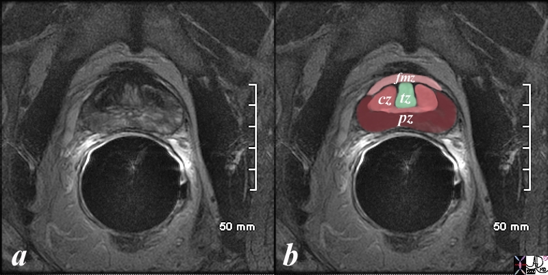
Normal Zonal Anatomy MRI T2 Weighted |
|
This image was described above in the “character” section but is repeated in the context of the parts of the gland. PZ = peripheral zone, CZ = central zone, tz = transitional zone and fmz = fibromuscular zone or fibromuscular layer Image Courtesy Ashley Davidoff MD Copyright 2010 98877c.8s |
The prostate contributes to both the structure of the internal and external sphincter. The anterior fibromuscular layer contributes smooth muscle to the internal sphincter and skeletal muscle from the inferior aspect of the anterior fibromuscular zone, and is part of the external sphincter. The internal sphincter is under autonomic control while the external sphincter is under voluntary control.