
The Common Vein Copyright 2010

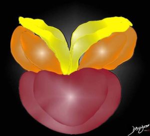
The artistic rendition is of the normal prostate enlarging with time most commonly due to hyperplasia of the transitional zone. The diagram shows the cone shaped gland in maroon with the ductus deferens (yellow) and seminal vesicles. The prostate is about the size of a kiwi fruit usually weighs between 20-30gms and has a maximum transverse diameter of 4cms.
Image Courtesy Ashley Davidoff MD Copyright 2010 99380b03b.8s
Introduction

|
|
The artistic rendition is of the normal prostate enlarging with time most commonly due to hyperplasia of the transitional zone. The diagram shows the cone shaped gland in maroon with the ductus deferens (yellow) and seminal vesicles. The prostate is about the size of a kiwi fruit usually weighs between 20-30gms and has a maximum transverse diameter of 4cms. Image Courtesy Ashley Davidoff MD Copyright 2010 99380b03b.8s |
 Getting Bigger
|
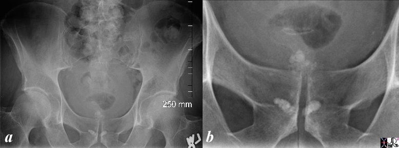
Dystrophic Calcification Above the Pubic Symphisis Indicates an Enlarged Gland 79 Year Old Male |
|
The plain film of the pelvis is from a 79 year old man. Two areas of coarse calcifications are noted in the midline. In this patient they have a linear appearance inferiorly and lobular appearance superiorly. These coarse calcifications are common and “normal” findings occurring in men after the age of 50 commonly as a result of dystrophic calcification of thickened secretions that occur with advancing age. These secretions are called corpora amylacea. The presence of the calcifications above the pubic symphisis confirm enlargement of the gland. Calcification of the prostate gland is most commonly caused by benign prostatic hypertrophy or chronic prostatitis. The calcifications can be identified at or below the pubic symphisis. When the gland is hypertrophied they may project above the symphysis. Calculi may form within the prostate gland in dilated prostatic or in Image Courtesy Ashley Davidoff MD Copyright 2010 98902cL.8s |
Normal – 26 year old male
|
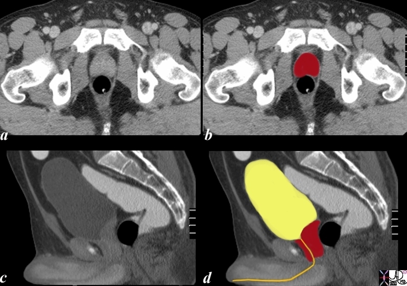
Normal Size Prostate 26 Year Old Male |
|
The CTscan is from a 26 year old male with a normal prostate seen in the axial plane (a,b) and in the sagittal planes (c,d). The bladder is distended. Note the shape of the gland in the transverse planer is heart shaped and in the sagittal plane almost rectangular when normal in size. The internal sphincter surrounds the top of the urethra at the base of the bladder and the urethra extends through the prostate as the prostatic urethra and into the penile urethra. Image Courtesy Ashley Davidoff MD Copyright 2010 99331c02.8s |
BPH
|
BPH |
|
The 70 year old male shows an enlarged prostate 7.2cms in A-P by 6.1cms. in the axial plane. The gland is enlarged based on these measurements. Two distinct zones are identified. . The posterior zone is canoe shaped is hypodense and represents the peripheral zone. The anterior more hyperdense layer is combination of zones. Since the prostate gland has 4 major zones and we have accounted for the peripheral zone (pz), three other zones are present. Inward of the peripheral zone is the central zone followed by the transitional zone and finally the anterior fibromuscular layer In this patient who has benign prostatic hypertrophy (BPH) the transitional zone is enlarged due to hyperplasia. Note that BPH is a misnomer because the enlargement is due to hyperplasia and not hypertrophy. Image Courtesy Ashley Davidoff MD Copyright 2010 25078c04L.8s |

|
BPH and Zonal Anatomy |
|
The 70 year old male shows an enlarged prostate 7.2cms in A-P by 6.1cms. in the axial plane. The gland is enlarged based on these measurements. Two distinct zones are identified. . The posterior zone is canoe shaped is hypodense and represents the peripheral zone. The anterior more hyperdense layer is combination of zones. Since the prostate gland has 4 major zones and we have accounted for the peripheral zone (pz), three other zones are present. Inward of the peripheral zone is the central zone followed by the transitional zone and finally the anterior fibromuscular layer In this patient who has benign prostatic hypertrophy (BPH) the transitional zone is enlarged due to hyperplasia. Note that BPH is a misnomer because the enlargement is due to hyperplasia and not hypertrophy. Image (c) illustrates the enlarged transitional zone which is where the hyperplasia of BPH takes place. Image Courtesy Ashley Davidoff MD Copyright 2010 25078c04bL01.8s |

|
Large Bilobed Smooth Gland |
|
The CTscan is from a patient with an enlarged prostate. The anterior aspect of the gland protrudes with two symmetrical homogeneous components likely representing an enlarged transitional zone and prostate hyperplasia though by the nature of the disease the combination of cancer and BPH commonly coexist. The urinary bladder wall is minimally thickened. Image Courtesy Ashley Davidoff MD Copyright 2010 25517b.8s |

|
Large Irregular Gland |
|
The CTscan is from a patient with an enlarged prostate. The anterior aspect of the gland protrudes into the bladder with an irregular shape (green arrow). The bladder wall is slightly thickened (orange arrow). The peripheral zone is suggested by a hypodense posterior region (maroon arrow) Image Courtesy Ashley Davidoff MD Copyright 2010 24842.81s |

|
Large Gland in the Sagittal Plane |
|
The CTscan reconstructed in the sagittal plane is from a patient with an enlarged prostate. The anterior aspect of the gland protrudes with two symmetrical homogeneous components likely representing an enlarged transitional zone and prostate hyperplasia though by the nature of the disease the combination of cancer and BPH commonly coexist. The matrix of the gland is heterogeneous The bladder wall is thickened anteriorly. Image Courtesy Ashley Davidoff MD Copyright 2010 76771.8s |
Carcinoma
|
Irregular Shape of Carcinoma as it Invades into Surrounding Tissue |
|
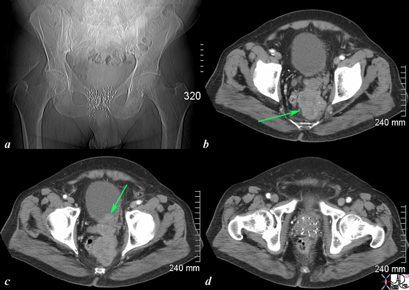
The CT scan of the pelvis is from an 76 year old man with known prostate carcinoma treated with implanted radiation seeds. Image a shows multiple radiation seeds within the prostate gland Image b (light green arrow) shows an irregular heterogeneous mass measuring about 5cms extending above the treated field and progressing posteriorly to the presacral space. . Image c shows the same mass as b extending into the base of the bladder (green arrow). Image d shows multiple prostatic seeds in the prostate gland. These finding are consistent with prostate carcinoma treated with implanted radiation seeds but with the tumor now continuing to grow superiorly beyond the confines of the treatment, and inward to the base of the bladder. . Image Courtesy Ashley Davidoff MD Copyright 2010 98929c01.8s |
|
Local Extensive Spread to Bladder and Hydronephrosis |
|
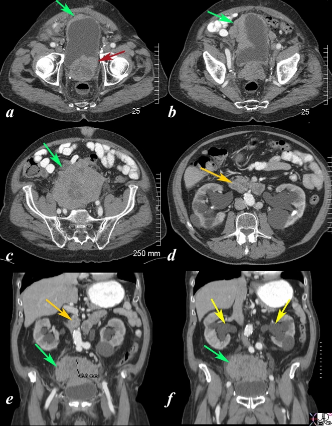
The CT scan is from a 88year old man with known prostate carcinoma. Image a shows multiple the mildly enlarged prostate (maroon arrow) with irregular margins extending to the left of the bladder but with a thickened right anterolateral wall (green arrow). Image b shows extensive irregularity of the superolateral wall which reflects tumor growth superior to and rightward of the bladder (see images below) (green arrow) . . Image c,e,f shows the large mass (green arrow) which has its epicenter above the bladder. Image d and e shows lymphadenoparthy (orange arrows ) anterior to the aorta resulting in hydronephrosis (yellow arrows in f. These findings are consistent with metastatic prostate carcinoma with extensive local extension above the bladder and to retroperitoneal nodes complicated by hydronephrosis. Courtesy Ashley Davidoff MD Copyright 2010 98999cL.81s |
|
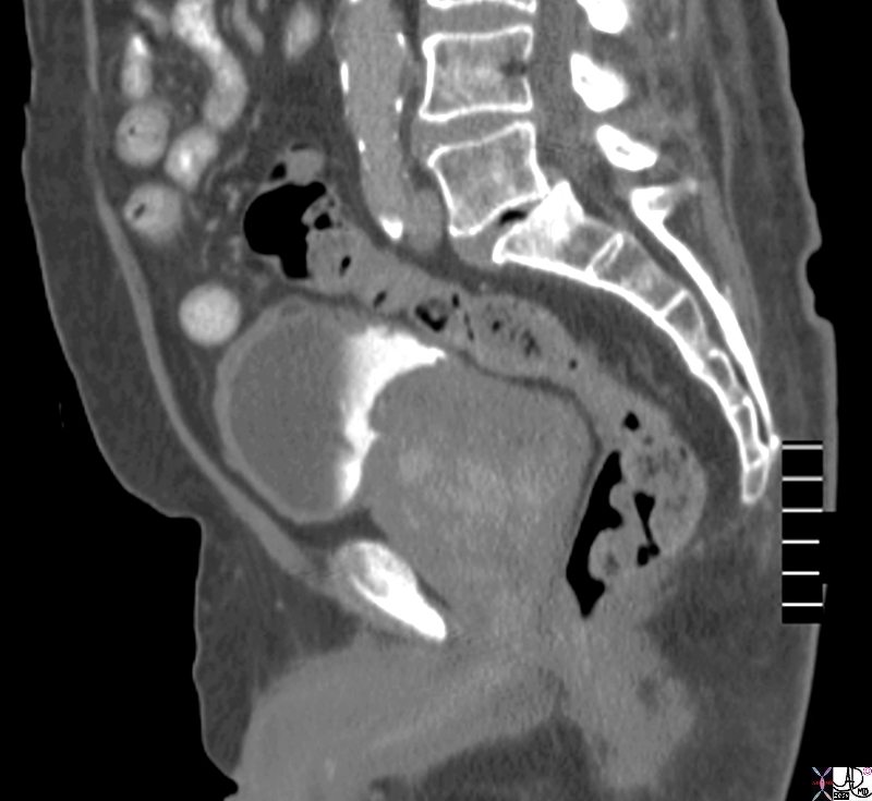
Obstructio and Hematuria |
|
The CT scans show an enlarged prostate (a) with hydroureters (b,c yellow arrow) blood-urine level or sediment urine level (red arrow c) with mild hydronephrosis (yellow arrows d) These findings are consistent with obstruction of the kidneys as a result of prostatic enlargement, and in this case prostatic carcinoma. Image Courtesy Ashley Davidoff MD Copyright 2010 18670cL.8s |
